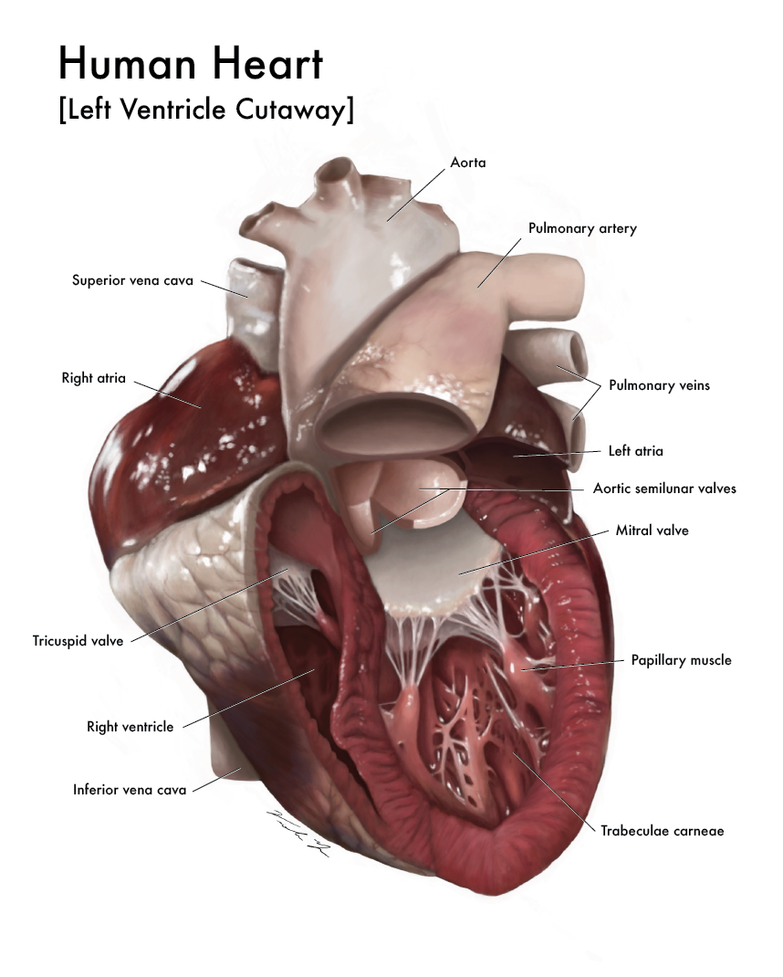
A Look Inside the Left Ventricle of the Human Heart
The goal of this illustration was to create an unique
educational illustration that informs the viewer of important external and
internal anatomical features of the human heart.
Continue reading to learn about the process that went into creating this piece.
Observational Sketches
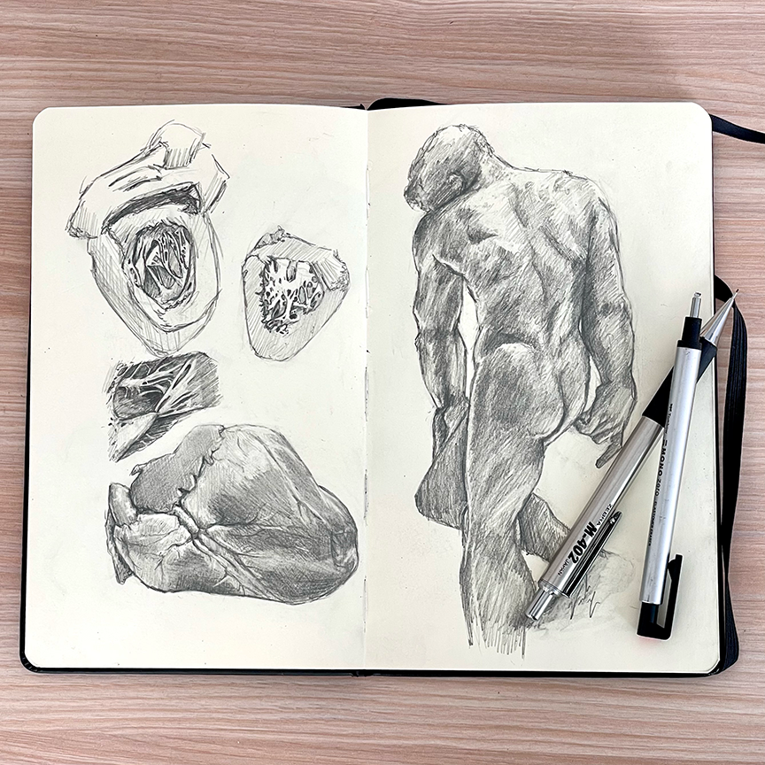
The first step after research into the anatomical information, is to create observational sketches. Top three sketches on the left page shows sketches done from life observing a human cadavear heart. With detail texture studies of trabeculae carneae and chordae tendineae. The bottom heart on the left page is a study of a pig heart.

Color Study
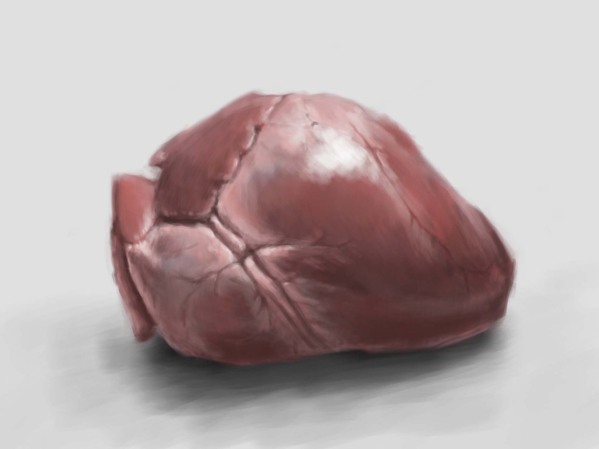
Graphite sketches while a good tool to explore the physical structures of the heart, does not give any information on chroma. Therefore, I decided to do a color study of a pig heart done as a digital painting. This gave my important information such as color undertones.

3D Maquette
The next step was to use a 3D maquette to compose the final illustration. The parameters of the assignment was to create a cut view in the human body that is atypical. Therefore I used this 3D model in Nomad Sculpt and created cut-outs with Boolean. After experimenting with different cuts, I settled on one that focused on the left ventricle.
Final Render
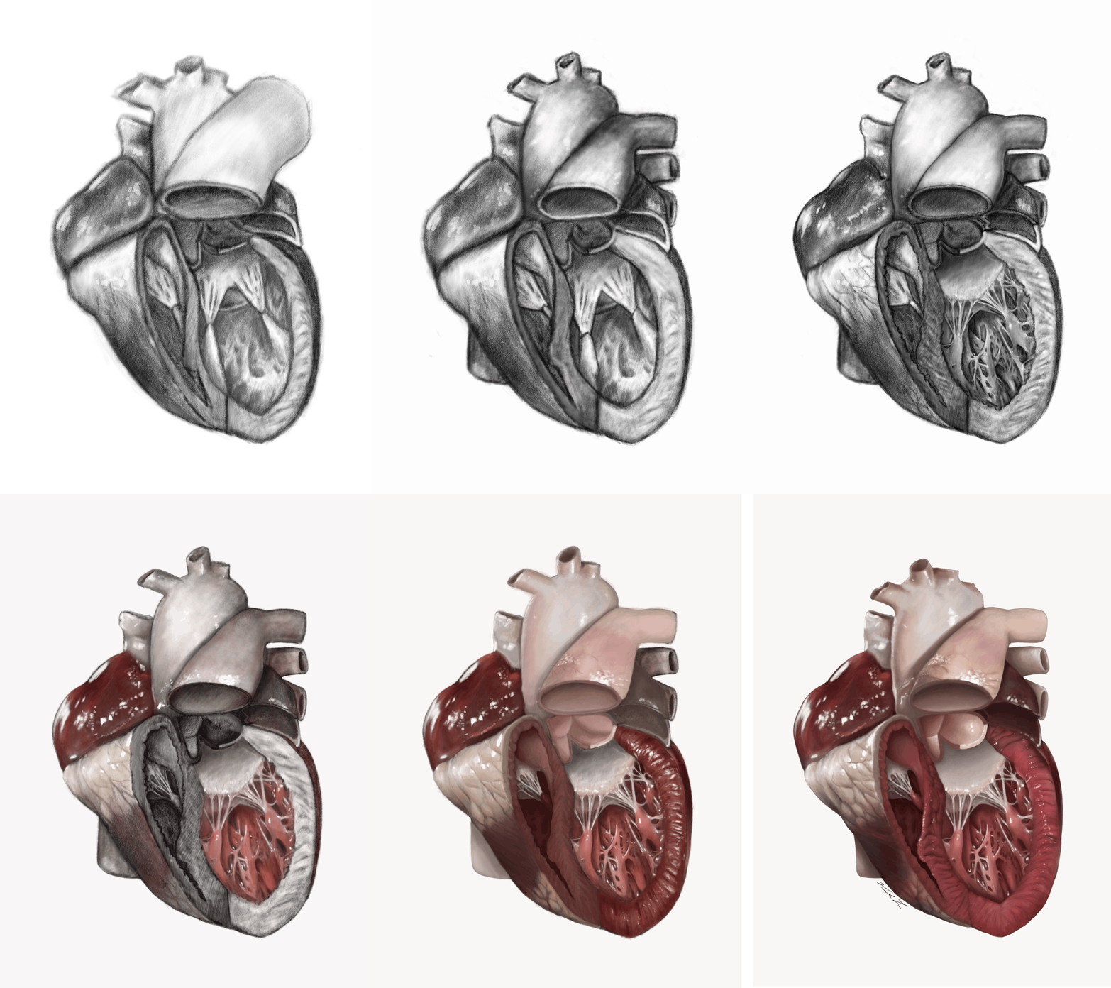
After exploring the physical structures of the heart, the coloration of the heart, and different cut-aways, the final render can start. With the physical structure of the heart being first rendered in black and white then color is layed on top.
